After
two long semesters at the University in Krems I was glad that summer
break had begun. One thing was clear: I wanted to take one month to
concentrate only on training. The plan was to focus on Kickboxing,
but I totally fell in love with Luta Livre and spent most of my time
learning about positions, escapes, passes and submissions. Soon I
stumbled upon a technique called „Heel Hook“, a submission which
attacks the anterior cruciate ligament (ACL). Pretty complicated stuff, so
while at class I was trying to understand how to actually do and
defend against this submission, after class I was part of some
interesting discussions about the cruciate ligaments. Eventually I
realized that I may not know as much about them as I thought I would.
Therefore, I wanted to do a blog post about the knee joint, focusing
on the cruciate ligaments, because I had found some interesting
things during my research I didn't know before.
 |
| click to zoom |
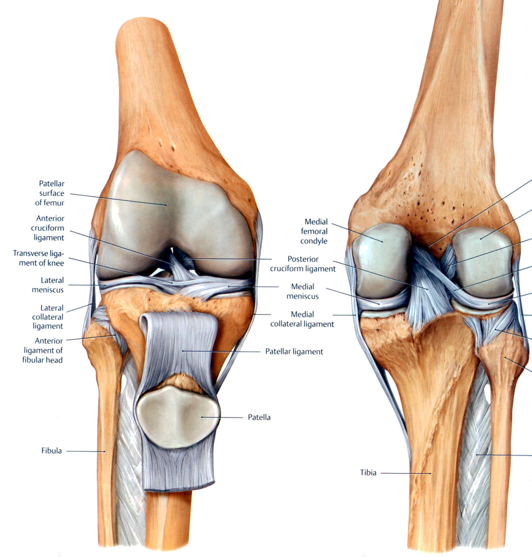
So
let's start with some basic anatomy of the knee: this joint is made
up of three bones, the femur, the tibia and the patella. Between the
tibia and the femur there are two „shockabsorbers“: the medial
and lateral meniscus. They transfer the load from the upper leg to
the lower leg and stabilize the knee. As you can see on the picture,
the femur and the tibia have different forms. The mensicii build a
link between those two bones so that they can fit together.
On
each side (medially and laterally) there is a ligament called
collateral ligament. The medial meniscus is grown together with the
medial collateral ligament, which makes injuries in this region more
complicated. The collateral ligaments give side way stability to the
joint. Between the two condyles of the femur and attaching to the
tibia, two ligaments cross each other – the anterior and posterior
cruciate ligament. Their function is not that simple, so later on I
will talk about them more detailed. But first let's talk about the
movement directions, because they are pretty simple. The knee can be
flexed and extended but can be rotated only very little.
Here
are two helpful videos, where you can get a visual impression of this
joint and it's characteristics:
 |
| click to zoom |
Although
at approximately 30 degrees of flexion neither of the bundles are
taught. This leads to the most translation available and is the
position where injuries in twisting movement most likely occur.
Generally speaking the problem with ligaments is, that they do not
stretch like tendons do. Imagine ligaments as ropes and tendons as
rubber bands for example. Stretching a tendon is part of everyday
life. But if a ligament gets stretched too much it tears apart.
 |
| click to zoom |
Let's
keep it simple and focus only on the ACL. Injuries of the ACL are
very common in the field of sports and much more likely than PCL
injuries. In sports cutting or sidestep maneuvers have a lot of
impact on the ACL. These movements can be found a lot in soccer games or american football for example. The typical injury occurs with the knee externally
rotated, in 10-30 degree flexion with the knee placed in a valgus
position. ACL tears also occur, if there is an anterior tibia
translation with the knee in shallow flexion.
Up
to 80% of the ACL injuries occur in non-contact situations, for
example if the athlete takes off with the aim of changing direction,
or in landing situations, with the knee close to full extension.
Especially landing situations require eccentric muscle action. If the
hamstrings and the quadriceps are weak, the tibia translates
anteriorly and the ACL might get injured.
The Heel Hook on the other hand is a little bit different, because it is a contact and not a non contact situation. During this
submission internal rotation on the tibia is applied via the heel.
Also in flexion the tibia acts as long lever arm that generates
increased forces to the joint. As we have heard before flexion
combined with internal rotational forces are very dangerous for the
ACL. So if a fighter finishes this submission and the opponent
doesn't tap in time, or the fighter is not cautious enough, the ACL
of the attacked fighter will be torn apart. Therefore, the Heel Hook
is known to be a very dangerous submission and is even forbidden in
some competitions. During training sessions I haven't had any
troubles with the Heel Hook, because if you know about its dangers
and are cautious in training sessions then injuries shouldn't happen
at all.

There are ways to prevent ACL injuries. A lot of exercises in the field of prevention are focusing on balance training, plyometrics, movement education and agility training. Next semester at University I will attend to a lecture about sport specific injury rehabilitation. Hopefully I will learn about this pathology and its rehabilitation a lot more, so I can tell you about it in another blog post.
Keine Kommentare:
Kommentar veröffentlichen